Interpreting the VPI MRI Study
Because 3D MRI allows for visualization of virtually any image plane, the types of measures that can be obtained are endless. This figure demonstrates a few of the common data points obtained in our laboratory studies using a (A) midsagittal (B) coronal (C) oblique coronal and (D) axial image plane.
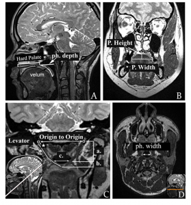
*Ph = pharyngeal; P = palate; Figure C(a) = extravelar levator veli palatini, C(b) = intravelar segment of the levator veli plalatini, C(c) intravelar distance
For clinical MRI studies, we tend to focus on the following:
- levator veli palatini muscle (morphology/shape and cohesiveness)
- relative position of the levator muscle (e.g., too close to the hard palate)
- pharyngeal depth
- velar length and thickness
- velopharyngeal gap (location, size, and type of closure patter)
- overall function of velopharyngeal structures and muscles
Overview of VP Anatomy
VPI-OPS will be the first study to determine the usefulness of these measures in assessing VPI and will evaluate which anatomic features are predictive of outcomes. Results from this study will be especially useful in advancing the potential of MRI in clinical VPI assessments.
How to Perform Measurements